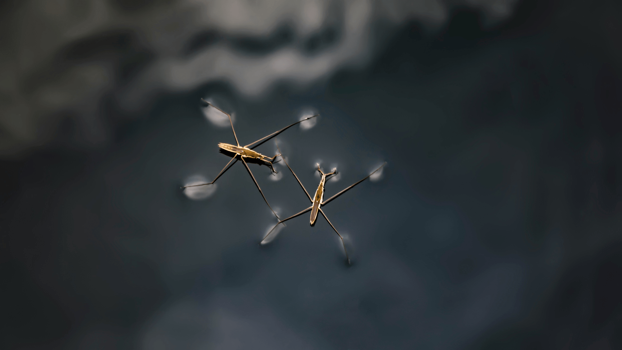
Tag: biology

  • Corralling Corals

    Corralling Corals

    So much of fluid dynamics is seeking patterns. Shown here are two sets of patterns, each created by a different species of coral larvae. These tiny creatures form a streaming flow (orange inset) around them as they swim. Combined together in a petri dish, the larvae follow winding paths, shown in white. The overall pattern is distinctly different for the two species. One shows a clear preference for paths near the wall of the dish (left), while the other corkscrews through open spaces (right). This difference raises questions researchers can explore: do the larvae differ in their propulsion methods or in their collective behavior? (Image credit: G. Juarez and D. Gysbers)

  • The Best of FYFD 2023

    The Best of FYFD 2023

    A fresh year means a look back at what was popular last year on FYFD. Usually, I give a numeric list of the top 10 posts, but this year the analytics weren’t as clear. So, instead, I’m combining from a few different sources and presenting an unordered list of some of the site’s most popular content. Here you go:

    I’m really pleased with the mix of topics this year; many of these topics are straight from research papers, and others are artists’ works. At least one is both. From swimming bacteria to star-birthing nebulas, fluid dynamics are everywhere!

    If you enjoy FYFD, please remember that it’s a reader-supported website. I don’t run ads and it’s been years since my last sponsored post. You can help support the site by becoming a patronmaking a one-time donationbuying some merch, or simply by sharing on social media. And if you find yourself struggling to remember to check the website, remember you can get FYFD in your inbox every two weeks with our newsletter. Happy New Year!

    (Image credits: sphinx – S. Boury et al., ear model – S. Kim et al., maze – S. Mould, dandelion – S. Chaudhry, water tank – P. Ammon, e. coli – R. Ran et al., drop impact – R. Sharma et al., Leidenfrost – L. Gledhill, toilet – J. Crimaldi et al., engine sim – N. Wimer et al., rivers – D. Coe, fin – F. Weston, snake – P. Schmid, nebula – J. Drudis and C. Sasse, flames – C. Almarcha et al.)

  • “The Reef”

    “The Reef”

    Artist Alberto Seveso returns to his colorful ink plumes (1, 2, 3, 4, 5), but this time with a twist. Here, Seveso took ink injected in water and digitally altered it, adding texture and shaping the ink to mimic the shapes of coral reefs. The results are stunning, though I confess a few of them remind me of mushrooms or organs more than reefs. (Image credit: A. Seveso; via Colossal)

  • Ciliary Pathlines

    Ciliary Pathlines

    For tiny creatures, swimming through water requires techniques very different than ours. Many, like this sea urchin larva, use hair-like cilia that they beat to push fluid near their bodies. The flows generated this way are beautiful and complex, as shown above. Importantly for the larva, the flows are asymmetric; that’s critical at these scales since any symmetric back-and-forth motion will keep the larva stuck in place. (Image credit: B. Shrestha et al.)

  • Featured Video Play Icon

    Inside a Zebrafish Heart

    This glimpse inside a 5-day-old zebrafish’s heart shows why they’re often used as a model organism in cardiac studies. The fish’s heart rate is similar to humans and its two-chamber heart — one atrium and one ventricle, both seen here — serves as a simplified version of ours. Check out the slowed-down section of the video to clearly see blood filling and expanding one chamber before it’s pumped onward. Perhaps the most unusual feature of the zebrafish’s heart is its ability to regenerate; after amputation of up to 20% of its ventricle, the fish can fully regenerate its heart. That’s a pretty incredible recovery, especially when you consider that the heart has to keep pumping the entire time! (Video credit: M. Weber/2023 Nikon Small World in Motion Competition)

  • Jamming Inside

    Jamming Inside

    Worm-like Spirostomum ambiguum are millimeter-sized single-cell organisms that live in brackish waters. In milliseconds, these cells can retract to half their original length, generating g-forces greater than a Formula One driver experiences when cornering. How, researchers wondered, do these cells avoid shredding their internal structure with forces that strong?

    Spirostomum ambiguum, they found, contain fluid-filled sacs called vacuoles that are entangled with the folds of a membrane-like structure called the endoplasmic reticulum. The researchers constructed a simulated cell, based on the properties of the living ones, and tested it under retraction. Without the endoplasmic reticulum, the insides of their model acted like a liquid, with vacuoles moving past one another readily. That’s not good for staying alive since swapping positions can disrupt bodily functions.

    An artificially-colored micrograph highlights the different structures inside Spirostomum ambiguum. The red strings are a membrane-like endoplasmic reticulum entangled between yellow, fluid-filled vacuoles.
    An artificially-colored micrograph highlights the different structures inside Spirostomum ambiguum. The red strings are a membrane-like endoplasmic reticulum entangled between yellow, fluid-filled vacuoles.

    With the vacuoles connected by a model endoplasmic reticulum, the cell’s insides acted more like a solid during retraction. The vacuoles deformed but fewer of them traded places, instead jamming together to prevent rearrangement. Mimicking this structure at a larger scale, the team suggests, could enable new types of shock absorbers. (Image and research credit: R. Chang and M. Prakash; via APS Physics)

  • A Better Ear Plug

    A Better Ear Plug

    Ear plugs can be wonderful at blocking outside noise, but they come with a downside: they typically amplify internal bodily sounds, like our heartbeat, breathing, and chewing. This effect, called occlusion, is distracting enough for some users to forego ear protection or hearing aids. But a new prototype offers a hope for an occlusion-free future without requiring active noise-cancelling.

    Most devices fit a short way inside our ear canals, which blocks outside sound well, but creates a little resonance chamber between the plug and our ear drums. It’s this gap that amplifies the low-frequency sounds within our bodies, making them seem much louder. To counter that, the team’s new plug contains foam sections arranged with hollow spaces between. By tuning the properties of the 3D-printed foam, they created a resonant structure inside the earbud that damps out those low-frequency body noises while still blocking outside sound.

    Illustration of the earbud's interior. The blue and green areas are foam-filled cavities.
    Illustration of the earbud’s interior. The blue and green areas are foam-filled cavities.

    So far the prototype has only been tested with an artificial ear designed for auditory tests; that’s enough to show that the concept works, but next they’ll redesign the bud to fit a human ear canal more comfortably. (Image and research credit: K. Carillo et al.; via APS Physics)

  • Understanding Cyanobacteria

    Understanding Cyanobacteria

    Over 2 billion years ago, cyanobacteria emerged as Earth’s first photosynthesizing organisms. Today they are widespread and critical contributors to both carbon and nitrogen cycles. Colonies can form large mats, like those pictured above, but, even at the microscale, cyanobacteria are actively forming patterns among individual bacteria. A recent study considers cyanobacteria as active matter.

    At the microscopic scale, cyanobacteria form different patterns.
    At the microscopic scale, cyanobacteria form different patterns, depending on their density.

    By simulating the cyanobacteria as filaments that interact through a series of simple rules, the researchers were able to reproduce the complex patterns bacterial colonies form. Their physical model also offered an explanation — based on the relative importance of advective and diffusive transport — for the characteristic length scales found in the bacterial patterns. (Image credit: Yellowstone – B. Cappellacci, patterns – M. Faluweki et al.; research credit: M. Faluweki et al.; via APS Physics)

  • Swimming Through Mud

    Swimming Through Mud

    At the bottom of ponds, nematodes and other creatures swim in a world of mud. They squirm their way through a sediment of dirt particles suspended in water. Mud, of course, is notoriously impossible to see through, so to understand these creatures’ movements, scientists turn instead to biorobotics. Here, a team uses a magnetic head attached to an elastic tail to mimic these tiny creatures.

    To drive the robot’s motion, they use an oscillating magnetic field, which forces the magnetic head to rotate. Combined with the elastic tail and the drag caused by surrounding materials, this causes the robot to swim in a fashion similar to its biological inspirations.

    A biomimetic robot swims through immersed grains. The robot's magnetic head is forced with an oscillating magnetic field. It swims through an underwater bed of hydrogel beads, whose diameter is smaller than that of the robot's head.
    A biomimetic robot swims through immersed grains. The robot’s magnetic head is forced with an oscillating magnetic field. It swims through an underwater bed of hydrogel beads, with diameters smaller than that of the robot’s head.

    To mimic the muddy environment of a pond’s bottom, scientists used a bed of hydrogel beads immersed in water. Looking at the experimental video above, you’ll see no sign of the beads. That’s because the hydrogel beads have nearly the same index of refraction as water. Once you pour water in, they seem to disappear. That allows the researchers to focus instead on the robot’s motion. In other experiments, they added dye to the beads so that they could see how they moved around the robot.

    They found that the robot’s motion fluidizes the grains around it. Effectively, the robot’s motion creates an area with fewer grains and more water for it to move through. Once it’s passed, however, more grains settle in, and the bed returns to a denser packing. (Image credit: nematode – P. Garcelon, experiment – A. Biswas et al.; research credit: A. Biswas et al.)

  • Imitating a Cough

    Imitating a Cough

    Coughing and sneezing create violent air flows in and around our bodies. As that fast air rushes over mucus layers in our lungs, throat, and sinuses, the resulting flow breaks up the mucus into droplets. To explore the details of that process, researchers built a “cough machine” that sends a rush of air over a thin film of water mixed with glycerol. The setup allows them to observe the physics in a way that’s nearly impossible in a human cough or sneeze.

    Imitating a cough: high-speed video shows how a thin film made of water and glycerol breaks down in a strong airflow. Parts of the film inflate into hollow bags that form thinner weak spots. When the film breaks in those places, it forms rims and ligaments that break up into droplets.
    Imitating a cough: high-speed video shows how a thin film made of water and glycerol breaks down in a strong airflow. Parts of the film inflate into hollow bags that form thinner weak spots. When the film breaks in those places, it forms rims and ligaments that create a spray of droplets.

    As seen above, air flowing past shears the viscous fluid, stretching it out. The leading edge of the film destabilizes and breaks into large drops, but it’s what comes next that really gets things going. Areas of the film inflate to form hollow bags. When sections of the bag thin to about 1 micron, the film ruptures and the bags burst. This triggers a cascade of instabilities in the film’s rim that ultimately rip the film into a spray of tiny aerosol droplets. The researchers found that, despite their tiny size, these droplets collectively carry a large volume of liquid, making them all the more important for understanding transmission of respiratory illnesses. (Image credit: top – A. Piacquadio, experiment – P. Kant et al.; research credit: P. Kant et al.)