Our ears, like those of many other animals, convert mechanical signals to electrical ones, through a Rube-Goldberg-esque series of transformations. External sound waves make their way down the soft tube of the ear canal, which funnels them to a thin-walled cone, the eardrum, that’s about half as large as a dime. Here, the vibrating air pushes against the cone’s membrane, and those vibrations travel onward through a linked trio of small bones that amplify the vibration’s amplitude.
The last of these bones presses against an even smaller, oval-shaped membrane. As the bone moves, it shakes the membrane, sending waves through the liquid on its other side. Those waves travel down the spirals of the tiny, pea-sized cochlea, named for a snail shell’s shape. As the waves move through the liquid, they bend bundles of hair-like strands back and forth, like tall grass waving in a breeze. The bending triggers a chemical that binds to nerves at the base of the bundles, sending an electrical signal through the nerve and into the brain.
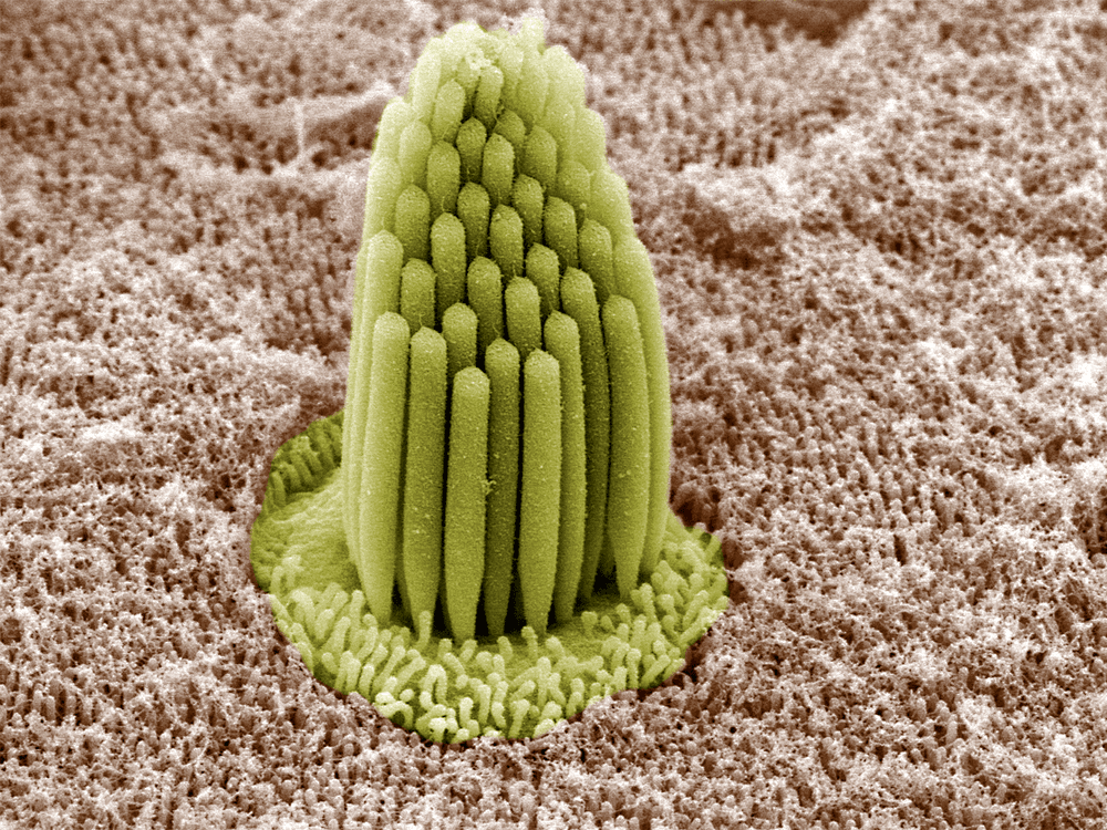
But the hair-like bundles, known as stereocilia, are also able to amplify incoming vibrations. In this case, the bundles in the outer portion of the cochlea expend energy to bend more than the incoming vibrations naturally make them move. This bending amplifies the fluid motion that gets transmitted to stereocilia further down the line; it’s those bundles that will make the final conversion to an electrical signal the brain receives. (Image credit: B. Kachar; research credit: Y. Thipmaungprom et al.; via APS)
Leave a Reply