Tag: placenta

  • Disease and Placental Flows

    Disease and Placental Flows

    The human placenta functions as a life-support system for a growing fetus. Despite its frisbee-like appearance, the organ is packed with nearly 10 square meters of blood vessels. On the fetal side, these blood vessels form villous trees where diffusion across the placental boundary exchanges molecules with the maternal blood that fills the space between villous trees. This setup allows oxygen, glucose, carbon dioxide and other key chemicals to cross between the parent and fetus while (ideally) keeping diseases out.

    Views of the placenta. Beige areas show the intervillous space where maternal blood flows while pink areas show villous trees where exchanges between the fetus and mother take place. The first three images show a) a preeclamptic, b) a normal, and c) a diabetic placenta. The final image d) shows a 3D view of placental tissue taken with x-ray tomography.
    Views of the placenta. Beige areas show the intervillous space where maternal blood flows while pink areas show villous trees where exchanges between the fetus and mother take place. The first three images show a) preeclamptic, b) normal, and c) diabetic placentas. The final image d) shows a 3D view of placental tissue taken with x-ray tomography.

    But when diseases directly affect the structure of the placenta, flow to the fetus gets disrupted. The image above shows cross-sections of placental tissues, with villous trees marked in pink, under (a) preeclamptic, (b) normal, and (c) diabetic conditions. Preeclampsia is associated with reduced density of villous trees, which restricts the amount of nutrients a fetus receives and can lead to reduced growth or stillbirth. In contrast, with gestational diabetes villous trees can proliferate, causing a high resistance to flow that also affects exchanges.

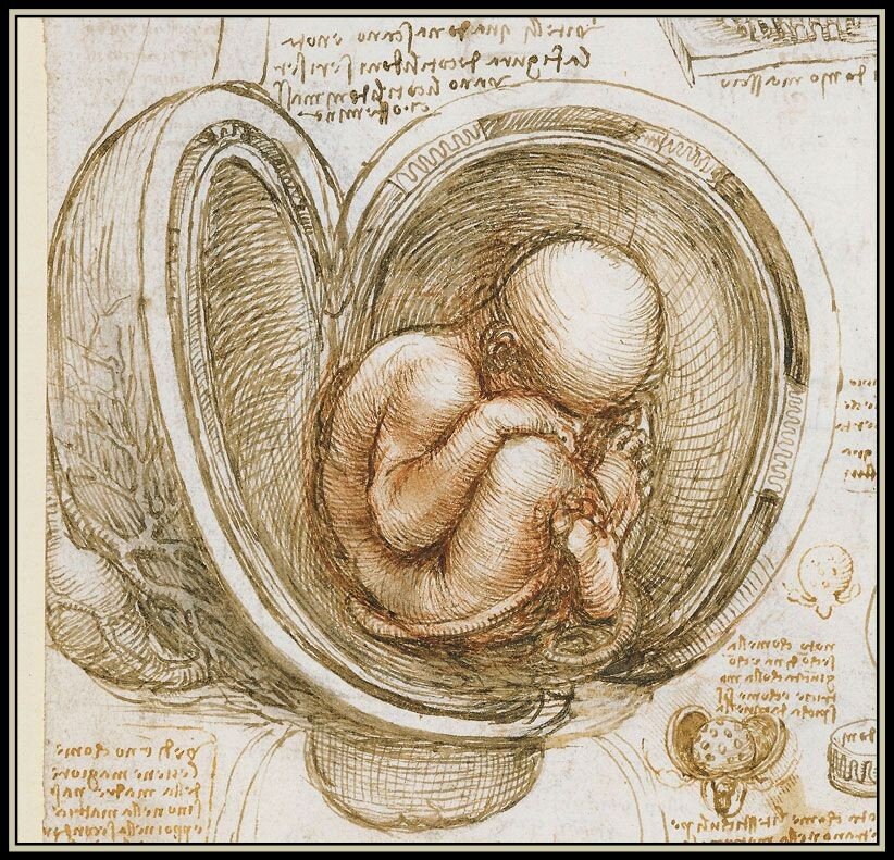
    For more on the complex physics of the placenta, check out this article from Physics Today. (Image credit: sketch – L. da Vinci, placentas – A. Clark et al.; see also A. Clark et al.)

  • Placental Fluid Dynamics

    Placental Fluid Dynamics

    The placenta, critical as it is to human life and development, is likely the least-studied organ in the body. Reasons for that abound, from the ethics of studying pregnant people to the historical marginalization of female bodies in medical studies. But what little we do know shows that the placenta is quite incredible.

    Shaped somewhat like a flattened cake, the placenta contains a tangle of fetal blood vessels — an estimated 550 kilometers’ worth — bathed in maternal blood. The enormous surface area — nearly 13 meters squared — enables the exchange of oxygen, glucose, and urea through diffusion. These exchanges don’t take place in still conditions, though; blood is always flowing through the vessel network. This means that each exchange depends on both the speed of diffusion and the speed of the flow, a relationship that’s captured with the dimensionless Damköhler number.

    Illustration of the intertwined blood vessels of the placenta.
    Illustration of the intertwined blood vessels of the placenta.

    Some exchanges, like carbon monoxide and glucose, are diffusion-limited, meaning that increased blood flow cannot speed up the process (though additional blood vessel surface area could). In contrast, carbon dioxide and urea are flow-limited exchanges. Fascinatingly, oxygen is close to being both diffusion- and flow-limited, suggesting that the organ has optimized for this exchange. Since pregnancy also involves a large increase in maternal blood volume and changes in lung capacity to help provide oxygen, it seems like the pregnant body heavily emphasizes delivering oxygen to the developing fetus. (Image credit: newborn – J. Borba, placenta – iStock/Sakurra; via Physics World; submitted by Kam-Yung Soh)